


A dental implant close to a nerve: what do you need to know before the procedure?
 Author: Dr. Bogdan Bumbu
 September 25, 2026
A dental implant placed in the posterior region of the mandible may be situated close to the mandibular canal, the bony structure through which the inferior alveolar nerve passes. The available space varies from patient to patient, which is why an assessment of the anatomy prior to the procedure is essential.
Proximity to the nerve does not automatically mean that the implant cannot be placed. It does, however, mean that its position and size must be carefully planned, depending on the available bone and the future restoration.
Before placing an implant close to the nerve, the dentist assesses:
- the distance from the mandibular canal;
- the height and thickness of the bone;
- the length and diameter of the implant;
- the position of the future crown;
- any alternatives if there is insufficient bone available.
What does it mean to have a dental implant close to a nerve?
The inferior alveolar nerve passes through the mandible via the mandibular canal and plays an important role in the sensation of the lower teeth; through its branches, it also contributes to sensation in the lower lip and chin.
In the posterior region of the mandible, implants can be planned relatively close to this canal. The available space depends on both individual anatomy and the amount of bone remaining following tooth loss.
Following extraction, the alveolar ridge undergoes a natural process of remodelling and may shrink in size. If bone loss is significant, the available height above the mandibular canal may be reduced.
This is why the same implant length is not automatically suitable for every patient.
What are the risks if the implant is too close to a nerve?
Injury to the inferior alveolar nerve
If the inferior alveolar nerve is affected during the procedure, changes in sensation may occur in the corresponding area.
The patient may experience:
- numbness in the lower lip or chin;
- tingling;
- reduced or altered sensation;
- a burning sensation;
- unusual discomfort or pain.
Symptoms may vary in intensity and duration, depending on the mechanism and extent of nerve damage. Numbness and paraesthesia are among the most commonly reported symptoms in nerve damage associated with mandibular implants.
Why is it important to prevent complications?
Some sensory disturbances may be temporary, but others may persist. For this reason, prevention is far more important than managing a complication once it has arisen.
Assessing the anatomy, identifying the mandibular canal and correctly selecting the position and size of the implant are all part of the planning stage.
What role does CBCT play in planning a dental implant?
What information does a CBCT provide?
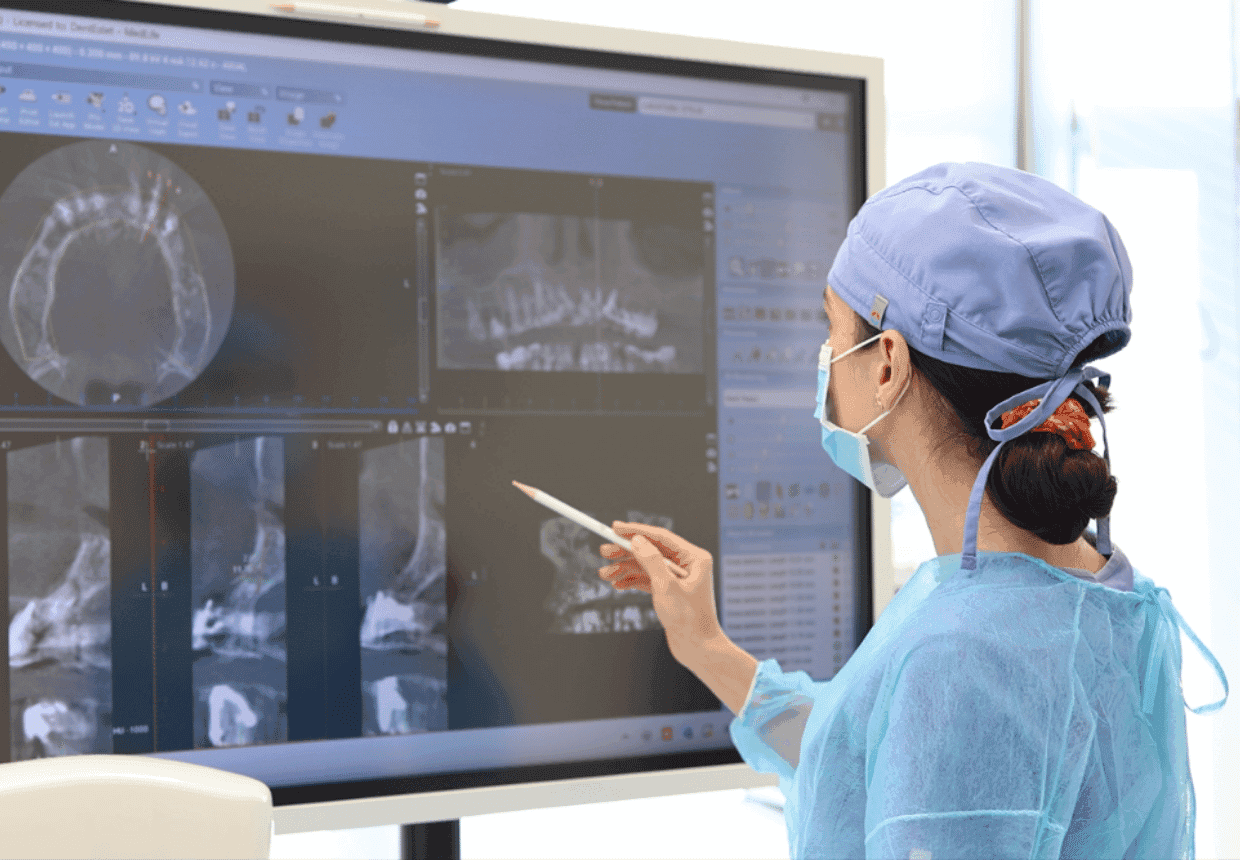
CBCT, or cone-beam computed tomography, provides three-dimensional information about the area where the implant is to be placed.
Where appropriate, the dentist can assess:
- the position and course of the mandibular canal;
- the height and thickness of the available bone;
- the shape of the alveolar ridge;
- the relationship between the implant site and neighbouring anatomical structures;
- any anatomical features relevant to the procedure.
The American Academy of Oral and Maxillofacial Radiology (AAOMR) recommends CBCT as the method of choice for pre-operative cross-sectional imaging of implant sites; the investigation is indicated on the basis of clinical assessment and with a field of view tailored to the area of interest.
Why is a panoramic X-ray not always sufficient?
A panoramic X-ray provides a two-dimensional image and can be useful for an initial general assessment. However, it does not allow for a three-dimensional assessment of bone thickness or the spatial relationship with the mandibular canal.
When this information is required to determine the position of the implant, CBCT provides important additional data for planning.
How do you plan an implant when the nerve is close by?
Planning begins by determining the position the implant must occupy in order to properly support the future crown.
Based on the clinical examination and imaging studies, the dentist determines the dimensions and position of the implant so that there is a favourable relationship with both the available bone and the surrounding anatomical structures.
The following are taken into account:
- the three-dimensional position of the implant;
- its length and diameter;
- the distance from the mandibular canal;
- the amount of available bone;
- the position of the future restoration;
- the possibility of using digital planning, where indicated.
The aim is not merely to avoid the mandibular canal. The implant must also be correctly positioned from a prosthetic point of view, so that the future restoration can function properly.
What options are available if there is limited bone available above the nerve?
Implants of customised sizes
In certain situations where bone height is limited, shorter implants may be considered.
However, these are not a one-size-fits-all solution. The choice of length and diameter must be made based on the patient’s anatomy, the treatment area, functional requirements and the type of restoration.
The ITI Consensus Guidelines consider short implants to be a valid option in certain cases involving reduced bone height, including where their use may reduce the risk of damaging anatomical structures such as nerves.
Choosing a different position
In certain cases, the position of the implant can be adjusted to make use of a more favourable bone area.
However, this adjustment must remain compatible with the position of the future crown. It is not advisable to simply move the implant to an area with more bone if this compromises the prosthetic outcome.
Other surgical options
Where the amount of available bone does not permit the insertion of an implant under appropriate conditions, other treatment options may be available.
Depending on the specific details of the case, bone augmentation procedures or other surgical approaches may be considered. The choice is made on a case-by-case basis, following clinical, imaging and prosthetic assessments.
Which post-implantation symptoms should be assessed by a doctor?
Following the procedure, there may be localised pain, tenderness and inflammation associated with the normal healing process.
However, changes in sensation must be monitored closely. If, after the anaesthetic has worn off, the following persist or occur:
- numbness in the lower lip or chin;
- tingling;
- a burning sensation;
- reduced or altered sensation;
- unusual pain;
- it is recommended that the patient contact their doctor for assessment.
Early diagnosis is important when nerve damage is suspected. The specialist literature emphasises the importance of prompt assessment when sensation remains altered after the anaesthesia has worn off.
What needs to be assessed before a mandibular implant?
Before treatment, the plan must be based on the patient’s actual anatomy, not simply on the location of the missing tooth.
The following are assessed in particular:
The distance to the mandibular canal
It is important to know the relationship between the area of the future implant and the course of the mandibular canal.
Available bone volume
The height and thickness of the alveolar ridge influence the size and position of the implant.
The position of the future implant
The implant must be planned in such a way that it respects the anatomy whilst also allowing for the correct fabrication of the future crown.
Implant size
The length and diameter are selected on a case-by-case basis. A longer implant is not automatically a ‘better’ implant if the anatomy does not allow it to be used under favourable conditions.
Available alternatives
If bone volume is limited, the dentist may consider implants of different sizes, a different positioning strategy or additional procedures, depending on the individual case.
Frequently asked questions about dental implants near a nerve
Can an implant be fitted if the nerve is very close by?
This may be possible, depending on the amount of available bone, the position of the mandibular canal and the feasibility of selecting a suitable implant size and position. The answer is determined on a case-by-case basis following clinical and imaging assessment.
What happens if the nerve is damaged during the procedure?
Changes in sensation, such as numbness, tingling or other unusual sensations in the lower lip or chin, may occur. Any such symptoms after the anaesthesia has worn off should be reported to your doctor.
Is numbness after an implant normal?
Numbness caused by local anaesthesia is normal whilst the anaesthetic is still taking effect. If the numbness persists after the anaesthetic should have worn off, or if you experience any unusual changes in sensation, you should seek medical advice.
Is a CBCT scan compulsory before having an implant?
The indication for the investigation is determined on the basis of the clinical situation. For three-dimensional planning of an implant site, and particularly when the relationship with the mandibular canal needs to be assessed accurately, CBCT can provide essential information.
Can shorter implants be used if there is insufficient bone?
In some cases, yes. Short implants may be an option when bone height is limited, but the decision to use them depends on a number of anatomical, surgical and prosthetic factors.
When should I contact my doctor after the procedure?
Contact your doctor if, once the anaesthetic has worn off, you notice persistent numbness, tingling, a burning sensation, unusual pain or any other changes in sensation.
Proper planning helps to minimise risks
“The proximity of the mandibular canal does not automatically mean that implant treatment cannot be carried out. It means that the anatomy must be carefully analysed and that the position and size of the implant must be determined prior to the procedure. Clinical assessment, three-dimensional imaging where indicated, and planning based on the future restoration all contribute to choosing a solution tailored to each patient.” – Dr. Bogdan Bumbu