
Interpreting Dental X-Rays
What is a Dental X-Ray?
A dental X-ray is a two-dimensional (2D) X-ray examination or digital scan of the oral cavity, resulting in a flat image of the teeth, jawbones, and surrounding soft tissues.
A dental X-ray is a non-invasive investigation that helps dentists make an accurate diagnosis and treat dental issues effectively. X-ray imaging involves exposing part of the body to a low dose of ionizing radiation, which is safe for the body.
The Role and Necessity of Dental X-Rays
Dental X-rays serve two main purposes. First, they provide answers to questions that a dentist may have when a regular exam does not reveal all of a patient’s dental issues. In cases where no visible signs appear on the teeth or gums, a dentist may need additional tools to reach a conclusion.
With an X-ray, the dentist can determine the nature of a treatment and even create a comprehensive treatment plan.
Additionally, X-rays play a preventive role, as they allow for the early detection of issues so that the appropriate treatment can be applied. Dentists need an accurate X-ray to identify cavities or other structures hidden beneath the surface of molars, early-stage wisdom teeth, bone loss, or any other conditions that are not visible to the naked eye.
How is a Dental X-Ray Taken?
X-rays are a type of electromagnetic radiation, similar to light or radio waves, that can penetrate body tissues, creating an image on a photographic film or specialized detector.
Most X-ray images are stored as digital files, which are easily accessible to the dentist for diagnosis and dental care management. The digital format also allows for adjustments in contrast and brightness, improving the visibility of specific structures and tissues.
Different tissues show up on X-rays in various shades, with harder tissues appearing lighter and softer tissues darker.
For example, bones or dental structures appear lighter on the radiographic image, while soft tissues such as the tongue, gums, or cheek mucosa appear as faint shadows.
Interpreting the Dental X-Ray
After an X-ray is taken, the dentist interprets it to aid in diagnosis:
- Bright white areas on an X-ray may represent a filling, crown, implant, or even an overlap of two or more teeth;
- Deep black areas can indicate a defect in hard structure (bone or tooth) or even its absence.
Identifying Pathological Conditions on a Dental X-Ray
Tooth Decay: Appears as a grey-to-black area on the tooth’s surface. Decay can also affect teeth with prosthodontic rehabilitations, typically at the junction between the natural tooth and the prosthesis.
Root Infections: Seen as mostly round grey or black areas at the root tips. If these areas are larger and well-defined, it could indicate a cyst or apical granuloma.

Bone Resorption: Shows up as a recession or elevation of the bone line below the normal level (where the natural tooth crown meets the root), exposing root areas surrounded by dark zones.
Large Cyst or Granuloma: Appears as an intense grey-to-black area originating from a tooth root and expanding. It should not be mistaken for the nasal area, maxillary sinuses, or lower mental foramina, which are normal anatomical structures.
Canines or Wisdom Teeth: Panoramic X-rays can show canines or wisdom teeth that are embedded in the bone, often angled differently from other teeth.
Dental X-rays can also reveal issues with dental implants, appearing as intense grey areas around the implant.
Types of Dental X-Rays
There are various types of dental X-rays, with digital X-rays becoming increasingly popular over traditional film X-rays. A study by the NIH, the U.S. government’s medical research agency, found that 90% of dentists in Belgium use digital imaging technology.

Thanks to modern technologies, dentists can now view detailed images of teeth, jaws, and tissues on a monitor for a more thorough analysis with greater precision. For patients, the main advantage of digital X-rays is that the entire process takes no longer than five minutes.
Main Types of Dental X-Rays:
- Periapical X-Ray: Captures the entire tooth, showing the crown, root, and surrounding bone.
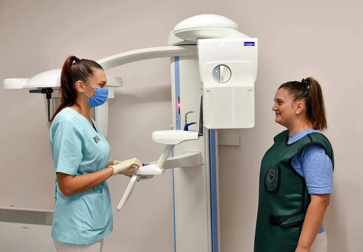
- Panoramic X-Ray: Shows both dental arches, including the maxillary sinuses and mandibular canals. The patient is fitted with a protective lead apron and positioned in front of the machine for the procedure.
- Occlusal X-Ray: Captures the entire dental arch along with the upper jaw and lower jaw. This type of X-ray is useful for detecting extra teeth, unerupted teeth, bone fractures, or foreign objects if the patient has experienced trauma.
- Bite-Wing X-Ray: An intraoral X-ray technique where the patient bites down on a device containing a small film. This type of X-ray shows the crowns of the posterior teeth in both the upper and lower jaw on the same image without overlapping.