
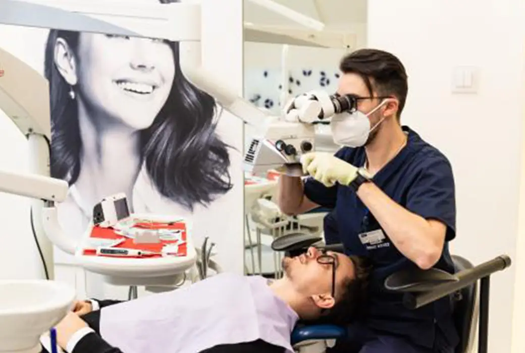
The microscope in dentistry
Redefinirea EndoExperienței
In realizing your treatment plan, your dental specialist chooses the optimal procedures to save your teeth and restore your long-term oral health. Modern endodontics allows visualization of the tooth's interior and its magnification to a level where it can provide the patient with highly predictable treatment solutions.
The possibilities offered by the dental microscope, in particular through its modern accessories, namely the intraoral camera, have made it possible to extend the limits of visualization of the inside of the tooth. This is why the dental microscope is used in many types of dental procedures, including endodontic surgery (apical resections and others).
In endodontics the dental microscope is used to save the tooth by treating the infection inside the tooth, in case of aggressive progression of untreated dental caries. This allows the endodontist to intervene with maximum precision and control over the affected area. We can even say that the endodontist performs a conservative procedure with the aim of protecting healthy tissue and preserving as much of the tooth's natural structure as possible.
More and more endodontists are opting to treat root canal fillings (root canals) with the aid of a microscope because the success rate is 90%, whereas statistics show that the success rate for traditional root canal treatment is around 50-60%.
The microscope helps the dentist have a much better view of the affected area and to be in control during the procedure.
Dr. Vlad Platon, endodontic dentist at DENT ESTET, explains in detail what this type of procedure consists of, presenting all the advantages that the patient can enjoy if he or she chooses a treatment with the dental microscope:
"First of all, the microscope ensures precision during the procedure and improves the safety of the work performed. Another advantage for the patient is that even the smallest carious processes can be treated and the intervention time is shortened. The microscope enlarges the field of vision and helps us see details of the tooth structure that we cannot see with the naked eye", remarks Dr. Vlad Platon.
The advantages of the dental microscope can be seen in the treatment of several conditions:
In the case of physiognomic fillings, up to 100% of the affected tooth tissue can be removed. The microscope gives the clinician a much better view of the interdental spaces, which helps us assemble a correct physiognomic filling in direct contact with the healthy dental tissue of the tooth.
Also, modelling the physiognomic filling with the microscope ensures that the future restoration of the tooth with a physiognomic filling is both aesthetic and, most importantly, functional.
Dr. Platon also adds, "The dental microscope helps us remove decay and protect the pulp chamber. Through the magnification of the image and the brightness it gives us, we have the necessary visibility to strictly remove the affected tissue."
Dr. Vlad Platon is one of the endodontic dentists in Romania who has specialized in the use of the microscope for both endodontic procedures and aesthetic conservative work.
In the DENT ESTET clinics, patients can choose Zeiss or Leica microscope treatment for precise and safe procedures, whether we are talking about physiognomic fillings, root canal treatments or endodontic surgery, such as apical resections.