
Digital Radiology Centre
Integrated digital radiology centre – what it is and how it helps patients
A digital radiology centre involves bringing together all the latest methods and technologies used in the diagnosis of dental conditions in a single location.
This means that the patient does not need to travel to another clinic for a radiological examination, and the dentist will receive the computer-processed three-dimensional image within minutes, enabling them to provide an accurate diagnosis and propose a treatment plan right from the first appointment.
What is digital dental X-ray?
Digital dental radiography is extremely useful in diagnosing dental conditions, providing the dentist with a highly precise tool for detailed imaging and measurements, on the basis of which the dentist can assess the quantity and quality of bone structure and visualise anatomical structures such as nerves or the maxillary sinuses.
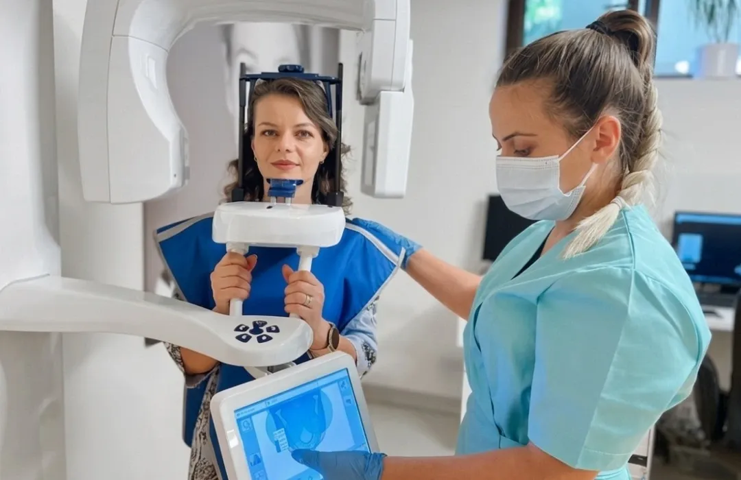
To produce a digital dental X-ray, a device is used that emits an extremely small amount of radiation, which captures the relevant information (teeth, roots, their position in the dental arch, bone) and transfers it immediately in digital format to a computer.
In this way, the dentist can observe, in real time and in detail, every dental structure—both bony and non-bony—and can provide the patient with an accurate diagnosis and a personalised treatment plan.
Key benefits of digital dental X-rays:
Accuracy in diagnosis
High-quality digital images
Minimal exposure to radiation
A patient-friendly procedure
Accuracy in identifying dental problems
Integration into digital treatment planning
More effective communication with patients
Storage and quick access to medical records
Types of digital X-rays
Digital dental X-rays can be taken intraorally and extraorally.
Intraoral digital dental radiography is the most common type of dental X-ray taken when diagnosing an oral condition and is used to identify dental caries and to check the condition and health of the teeth and jawbones.
Types of intraoral dental X-rays:
- Bitewing intraoral dental X-rays, taken by the patient biting down on film, provide details of the teeth in the upper and lower arches within a single area of the oral cavity. This type of digital X-ray is used to identify any interdental caries (between the teeth) and changes in bone density caused by periodontal disease. It also helps to check the fit of a dental crown following a prosthetic restoration and the integrity of the tooth following a filling.
- Intraoral periapical dental radiography captures the entire tooth on digital film, from the crown down to the root and the supporting bone tissues. Periapical dental radiography helps us identify abnormalities in the root and bone structure of the tooth.
- Extraoral dental radiography is used to identify affected teeth, to monitor the development of the mandible and maxilla, and to identify certain dental problems that may affect the temporomandibular joints or other facial bones.
Types of extraoral dental X-rays:
- Panoramic dental X-ray (OPG) – provides a digital image of the entire oral cavity, including the teeth of the upper and lower dental arches. It is used in dental implant treatments, identifies the position of the wisdom teeth, provides the dentist with accurate information about the jawbones and bone density, and helps diagnose bone tumours and cysts in the oral cavity.
- Cephalometric projections – are used primarily by orthodontists; they show the structure of the entire head and assist in the examination of dental conditions related to the jaws.
- Computed tomography (CBCT) – provides three-dimensional computerised imaging of the internal structure of the bones and tissues within the oral cavity and the head.
Frequently asked questions about digital dental X-rays
What can a digital dental X-ray reveal?
A digital dental X-ray can identify tooth decay, infections, dental abnormalities and any bone tumours. It also provides information about the structure of the tooth and can highlight problems with the temporomandibular joints.
What are the advantages of digital dental X-rays?
Digital dental X-rays enable the identification of sensitive areas, the creation of surgical guides and the personalisation of treatments, contributing to better communication with the patient and more predictable outcomes.
How long does it take to have a digital X-ray?
Taking a digital X-ray takes very little time, ranging from a few seconds to a maximum of 60 seconds, depending on the area being examined and the patient’s preparation.
Are there any risks or restrictions associated with radiological examinations?
Dental radiological examinations are safe and involve a very low level of radiation, particularly with digital technologies. They are only recommended by the dentist when necessary to establish an accurate diagnosis. In certain situations, such as pregnancy, the dentist will assess the appropriateness of the examination and take additional protective measures. The CBCT scanner emits the lowest level of radiation to which a person is exposed during a normal day’s activities or during a 3-hour flight (to London, for example).