
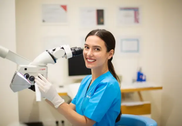
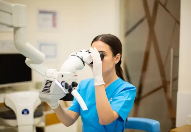
Zeiss or Leica Endodontic Microscopes
What isa dental microscope?
Many important elements of your oral cavity are microscopic in size. Special instruments are required for doctors to work with them. Since 1998, the dental microscope has become a key instrument in endodontics, and over the last 20 years it has also been adopted by other dental specialties such as periodontology and surgery. This dental microscope is designed for detailed visualisation during patient treatment.
Advantagesof using a dental microscope
The dental microscope offers visual accuracy 400 times greater than the naked eye and 100 times greater than traditional magnifying glasses. Magnification improves the accuracy of tooth preparation and prevents damage to neighbouring teeth and tissues during various surgical procedures.
In restorative dentistry, the use of a dental microscope increases visibility in diagnosis, preparation, execution and finishing of work. Dental microscopes have built-in high-intensity lights, which allow high visibility in areas that are difficult to access with the naked eye.
Many dental lesions, such as some types of cracks, are only visible with significant magnification, at least 16 times. Although many dentists limit themselves to using low-magnification or no magnifying glasses at all during examinations, dentists who work without a dental microscope are more likely to overlook small areas of decay.
A dental microscope is an invaluable resource for an endodontist. Because endodontic (root canal) treatment takes place in a very small area, the procedure requires precision to navigate the complex roots and canals. Missed dental canals often mean untreated infections, which can lead to repeat endodontic procedures.
At DENT ESTET clinics, we use the Zeiss dental microscope and the Leica dental microscope.
The dental microscope is used for:
Locating hidden dental canals that have been obstructed by calcifications and reduced in size.
Removing materials such as old filling materials.
Removing obstructions from dental canals.
Preparing access to avoid unnecessary tissue destruction.
Repairing perforations.
Locating cracks and fractures that are not visible to the naked eye.
Facilitating all aspects of endodontic surgery.
Improved photographic documentation.
Progress in dental microscope technology
The integration or attachment of a video camera to the dental microscope allows dentists to involve patients in their treatment more than ever. This significantly improves patient awareness, as their dental condition can be illustrated live. A clear, magnified image is worth more than a thousand words.
In the last three years, the use of dental microscopes has doubled. Dentists are constantly learning about dental microscope technology as it evolves rapidly. Since the 1990s, training in the use of dental microscopes has become an important component of endodontists' education. The use of dental microscopes is now universally taught to graduates in accredited endodontic specialty programs.