
Microscope-Assisted Dental Fillings
Advanced treatment for dental caries
Dental caries is the most common non-communicable disease globally, affecting over 2.5 billion people according to the World Health Organisation (WHO). Even when oral hygiene appears to be adequate, caries can develop and, if ignored, can quickly progress to more serious complications.
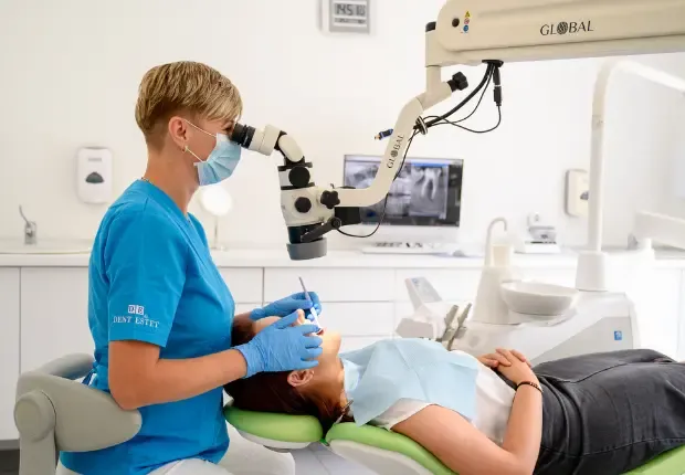
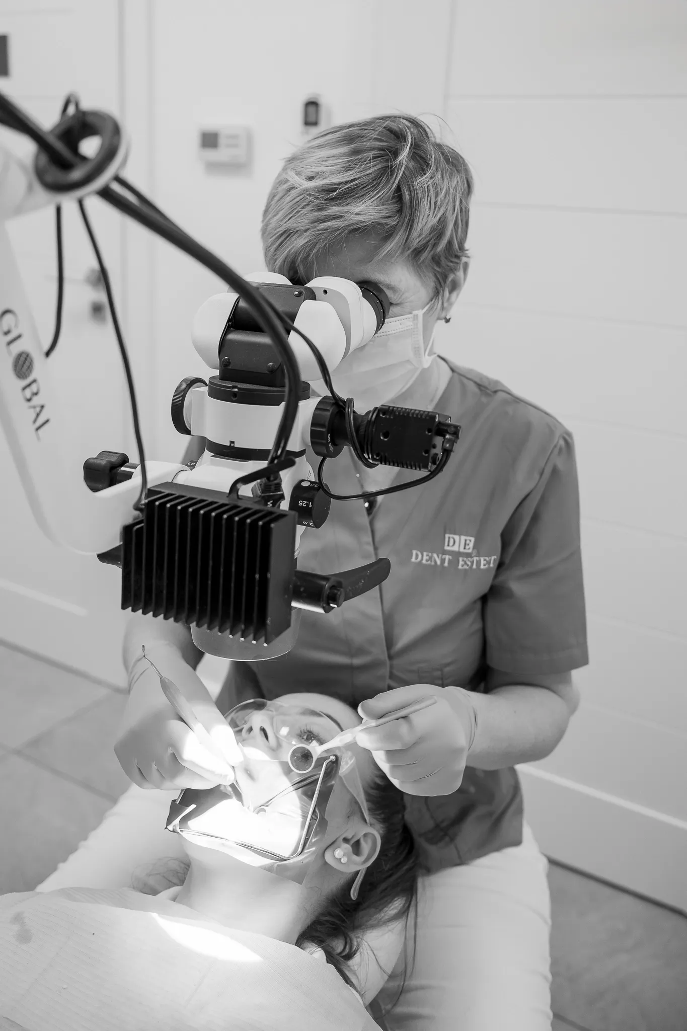
At DENT ESTET, we use a dental microscope to detect and treat cavities in their early stages, with a precision that is impossible to achieve with the naked eye. According to the American Association of Endodontists (AAE), the use of a microscope increases the success rate of treatments by visualising details that are invisible in conventional dentistry.
With this technology, our doctors are able to remove only the affected tissue, preserve the natural structure of the tooth and apply aesthetic, perfectly integrated fillings. The result? A healthy and natural smile, with minimally invasive treatments that are much more precise and durable, in accordance with the recommendations of the European Society of Endodontology (ESE) on the use of modern optical technologies.
Stages of treatment with a microscope
Accurate diagnosis
Before starting treatment, your dentist will recommend a series of tests, including a panoramic X-ray. They will then carry out a detailed examination using a state-of-the-art dental microscope. This allows us to accurately identify and assess any cavities and determine the appropriate treatment plan.
Tooth preparation
After diagnosis, the affected area is prepared for filling. Using the microscope, our dentist can effectively access and clean the affected area, completely removing the decayed tissue and preparing the surface for filling with the filling material.
Applying the filling
Once the area is prepared, our dentist precisely applies the filling material. This material is selected to perfectly match the colour and shape of the tooth, ensuring its aesthetic and functional reconstruction.
Adjustment and finishing
Finally, the filling is adapted to the occlusal surface so that it integrates perfectly into the bite, and then finished to blend in perfectly and restore the natural appearance of the tooth.
Benefits of microscope treatment for the patient
Early detection of incipient caries
According to the World Health Organisation, untreated cavities are the leading cause of tooth loss worldwide. The dental microscope allows intervention before the problem becomes serious.
Maximum preservation of healthy tissue
A study published in the Journal of Conservative Dentistry shows that using a microscope reduces the amount of healthy dental tissue removed by up to 30% compared to conventional treatments.
Impeccable aesthetics
Fillings made with modern composite materials, under microscopic control, integrate naturally and maintain colour stability for longer, in accordance with the recommendations of the European Society of Endodontology (ESE).
Reducing the risk of reoffending
According to the American Association of Endodontists (AAE), viewing microscopic details significantly reduces the chances of residual caries or imperfect fillings. The success rate of treatments increases from approximately 70% to over 90% when using a microscope.
Increased durability
A clinical study published in the International Journal of Microsurgery and Endodontics shows that fillings performed under a microscope have a 40% longer lifespan compared to fillings performed using traditional techniques.
At DENT ESTET, we ensure that each patient receives a personalised treatment plan. Depending on the complexity of the cavity and its stage of development, the dentist may recommend other types of interventions:
- For deep cavities that affect the dental nerve, the solution is root canal treatment under a microscope.
- For prevention and long-term oral health, regular dental prophylaxis is essential to remove plaque and tartar, the main causes of tooth decay and periodontal disease.
- The treatment is performed by the DENT ESTET team of doctors, who are experienced in using the latest technologies.
- See the complete list of DENT ESTET clinics throughout the country to choose the nearest location and schedule a consultation.
Frequently asked questions
Why is a filling done with a microscope better than a traditional one?
Because the dental microscope offers much better visibility and allows for the complete removal of decay, even from hard-to-reach areas. This preserves more healthy tissue and reduces the risk of the problem recurring.
How long does the procedure take?
The duration varies between 30 and 60 minutes, depending on the size and position of the decay. In the case of early decay, treatment is usually very quick.
What type of materials are used for fillings? State-of-the-art composite
materials are used, which faithfully reproduce the colour and transparency of natural teeth, ensuring an aesthetic and durable result.
Is the treatment painful?
The procedure is performed under digital anaesthesia, so the patient feels no pain. Thanks to modern technology, discomfort is minimal even after the treatment is complete.
How long does a filling made with a microscope last?
The lifespan is significantly increased compared to a filling made without a microscope, due to the precision and perfect integration of the material. With proper oral hygiene and regular check-ups, such a filling can last for many years.
Reviews
Marian Avram
July 22, 2026
Am pus de curând patru implanturi cu coroană fixă de zirconiu și sunt foarte mulțumit. Mulțumesc d-lor doctri Catalin Eremie si Alexandru Georgescu
Andrada Iacobescu
July 22, 2026
Cei mai buni!
Ciprian Scurtea
July 21, 2026
De câțiva ani apelez la serviciile clinicii Dent Estet Sibiu și sunt foarte mulțumit de modul profesionist și cald,în același timp, în care am fost tratat. Recomand clinica Dent Estet din tot sufletul!
Rancz Zsuzsa
July 21, 2026
profesionism amabilitate calitate punctualitate modern fără durere servicii excelente
violeta bianca Sirbu
July 16, 2026
Servicii de calitate,medici exceptionali, preturi lumesti, recomand din suflet ❤️ 10 stelute plus ❤️❤️
Ana Lita
July 16, 2026
Am avut o experiență foarte plăcută la Dent Estet Craiova . Recomand cu încredere pe doamna Dr. Dan Violeta.
Marilena Iacobescu
July 16, 2026
Foarte multumita de interventia dificila executata de Dnul dr Martisca Catalin si Dna dr Erika Vasilescu.
Roberta Badulescu
July 14, 2026
Am avut o experiență foarte plăcută alături de doamna doctor Dan Violeta. Este un medic profesionist, atent și explică fiecare etapă a tratamentului, ceea ce mi-a oferit încredere. Cabinetul este curat, modern, iar personalul este amabil și primitor. Recomand cu încredere tuturor celor care își doresc servicii stomatologice de calitate!”
Schedule a consultation
Fill out the form and one of our assistants will contact you to schedule a visit to the clinic.