
Interpretation of dental X-rays
What is a dental X-ray?
A dental X-ray is a two-dimensional (2D) X-ray examination or a digital scan of the oral cavity, producing a flat image of the teeth, the jawbones and the soft tissues.
An X-ray is a non-invasive examination that helps dentists make an accurate diagnosis and treat dental conditions. X-ray imaging involves exposing a part of the body to a small dose of ionising radiation, which does not pose a risk to health.
The role and necessity of dental X-rays
Dental X-rays serve a dual purpose. On the one hand, they provide answers to questions that dentists may have when a simple examination does not reveal all of the patient’s dental problems. Without obvious signs in the teeth or gums, the dentist needs additional tools to reach a conclusion.
With the help of an X-ray, the dentist can determine the nature of a dental procedure and even an entire treatment plan.
On the other hand, X-rays play a preventive role, as conditions can be detected early so that the appropriate treatment can be applied. The dentist needs an X-ray that is as accurate as possible to detect tooth decay or other structures invisible on the surface of the teeth, wisdom teeth in their early stages, bone loss or any other conditions that cannot be observed with the naked eye.
How is a dental X-ray taken?
X-rays are a form of electromagnetic radiation, just like light or radio waves. They have the ability to penetrate body tissues, ultimately producing an image on photographic film or a special detector.
Most X-ray images can be stored as digital files. These stored images are easily accessible to the dentist for diagnosis and management of dental conditions. The digital format also allows the dentist to adjust and change the contrast and brightness for a better view of specific structures and tissues.
Some of the scanned tissues are harder, whilst others are softer, which is directly reflected on the X-ray as lighter or darker areas.
For example, bones or dental structures appear lighter on the X-ray image, whilst soft tissues such as the tongue, gums or cheek lining appear as shadows that are harder to see.
Interpretation of dental X-rays
Once the X-ray has been taken, the dentist will proceed to interpret the dental X-ray for diagnostic purposes:
- The bright white area on the X-ray may represent a filling, a crown-root post, an implant, or even the overlap of two or more teeth;
- Deep black areas may indicate a defect in hard tissue (such as bone or tooth) or even its absence.
Interpretation of pathological conditions on dental X-rays
On an X-ray, dental caries appears as a grey to black area on the surface of the tooth. Caries can also affect teeth with dental restorations, right at the junction between the natural tooth structure and the restoration.
Infections at the root tips are shown as mainly round grey or black areas. If these areas are larger and more clearly defined, the diagnosis points towards a cyst or an apical granuloma.

Bone resorption is characterised by the bone line receding or advancing below the normal level (the boundary between the natural tooth crown and the root), leaving areas of exposed root surrounded by dark areas.
A large cyst or granuloma appears as an area ranging from deep grey to black, originating from a tooth root and extending into the surrounding area; This should not be confused with the nasal area, maxillary sinuses or inferior mental foramina, which are normal anatomical structures.
Canines or wisdom teeth appear on the panoramic X-ray, although they are not visible in the mouth. These are completely or partially embedded in the bone and, in most cases, are severely tilted relative to the other teeth.
Dental X-rays can also highlight problems with dental implants, which appear as areas of intense grey around the implant.
Types of dental X-rays
There are several types of dental X-rays, with digital X-rays becoming increasingly popular at the expense of traditional dental X-rays. A study published by the NIH, the US government’s medical research agency, shows that 90% of dentists in Belgium use digital imaging techniques.
With the help of the latest technologies, dentists can now view teeth, jaws and soft tissues on a computer screen, allowing for a more detailed analysis with a higher degree of precision. For the patient, the greatest advantage of digital radiography is that the entire procedure takes no longer than five minutes.
The main types of X-rays:
- A retroalveolar dental X-ray shows the tooth in its entirety. This reveals the crown, the root and even the bone in which it is embedded.
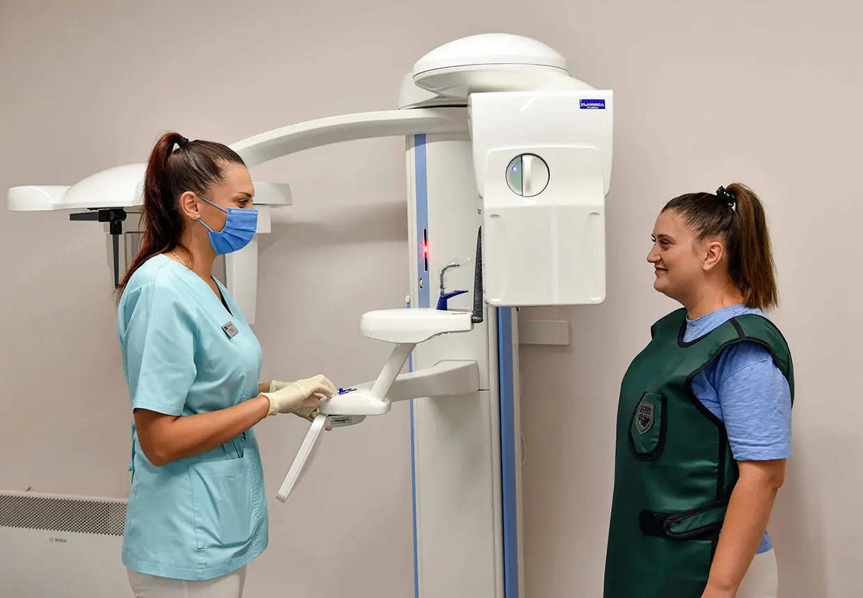
- A panoramic dental X-ray covers both dental arches, as well as the maxillary sinuses or mandibular canals. The procedure involves the patient wearing a lead apron and positioning themselves in front of the machine.
- An occlusal dental X-ray covers the entire dental arch, along with the upper jaw and lower jaw. The purpose of this X-ray is to detect supernumerary teeth, unerupted teeth, as well as bone fractures or foreign objects, should the patient visit the clinic following an injury.
- A bite-wing dental X-ray is an intraoral exposure. The distinctive feature of this type of dental X-ray is that the patient is asked to bite down on a device containing a small X-ray film. In this way, the crowns of the posterior teeth in the upper and lower jaws can be seen on the same X-ray, and the teeth are shown without overlapping.