
Microscope-Assisted Root Canal Treatment
Advanced precision for your dental health
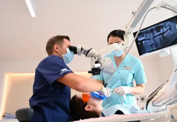
At DENT ESTET, we use the latest tech to give you effective, minimally invasive, and long-lasting treatments. Microscope root canal treatment is one of the ways we can save teeth that would have been pulled in the past. With a dental microscope, the endodontist can work with unmatched precision, clearing up the infection and restoring the tooth's function.
What is root canal treatment?
Root canal treatment (also known as root canal filling) is a dental procedure designed to save a tooth affected by deep infection or extensive decay that has reached the nerve (dental pulp). The procedure involves the complete removal of the inflamed or necrotic pulp, cleaning and disinfecting the root canal system and, finally, sealing it with biocompatible materials.
The aim of the treatment is to eliminate the infection, stop the spread of bacteria and preserve the natural tooth in the oral cavity, avoiding extraction.
This type of treatment is considered one of the most effective methods for saving compromised teeth, often being the only viable alternative before permanent tooth loss.
When performed correctly and completely – especially under a microscope – root canal treatment can provide stable and long-lasting results for many years.
When is root canal treatment necessary?
It is advisable to consult an endodontist if you have the following symptoms:
- Intense or throbbing pain in a tooth, especially at night
- Persistent sensitivity to hot or cold
- Inflammation or swelling in the gum area
- Discolouration of the tooth (usually turning grey or dark)
- Presence of pus or unpleasant taste in the mouth
- Dental trauma or deep cavities
A correct diagnosis, supported by X-rays, CT scans and microscopic evaluation, indicates whether root canal treatment is the right solution. These investigations are necessary to plan the procedure correctly or to decide whether a superficial cavity can be treated with simple fillings under a microscope.
Stages of root canal treatment under a microscope
Microscope root canal treatment is a procedure performed with greater precision than traditional methods, which are performed with the naked eye, due to the magnification and increased control over each stage. Here is how this treatment is performed at DENT ESTET clinics:
1. Diagnosis and planning
It all starts with a detailed assessment by the endodontist. This stage involves:
- Panoramic X-ray for an overview of the dental arch.
- Retroalveolar X-ray to analyse the structure of the roots and possible infections.
- CBCT (cone beam computed tomography) – 3D technology that allows three-dimensional analysis of root canals, hidden infections and possible anatomical complications.
- Microscopic examination to detect details invisible to the naked eye: microcracks, additional canals, perforations or previous treatments.
Based on this information, a personalised treatment plan is established, adapted to your dental morphology and the complexity of your case. The use of CBCT in planning is essential in complex cases, as it allows three-dimensional visualisation of the endodontic system, significantly reducing risks and increasing the precision of the intervention. This step helps us personalise the treatment and anticipate possible challenges, providing you with the safest and most effective treatment solution.
2. Root canal cleaning
This is one of the most critical stages of treatment:
- The dentist creates controlled access to the canal system using special rotary instruments.
- Under a microscope, all canals are identified – including accessory or atypical ones.
- With the help of antiseptic solutions and sonic/ultrasonic irrigation, the infection is completely eliminated.
- The dentist checks that no tissue debris, bacteria or fragments from previous treatments remain.
The use of a microscope significantly increases the chances of success and reduces the risk of reinfection.
3. Canal filling
Once all the canals have been cleaned and dried, they are sealed with biocompatible materials such as:
- Gutta-percha, an elastic and durable material
- Sealing cements, which complete the seal and prevent any bacterial penetration
The filling is done three-dimensionally, perfectly following the shape of the canal, to ensure a complete and durable treatment. The microscope allows the quality of the filling to be checked in real time.
4. Aesthetic and functional restoration
An endodontically treated tooth, even if saved by a correctly performed root canal treatment, may lose some of its strength over time. This is due to both the loss of tooth substance caused by deep caries and the dehydration of the tooth tissue with the removal of the pulp. Thus, post-treatment restoration becomes an essential step in protecting the tooth in the long term.
After closing the canals, the tooth must be restored so that it can withstand masticatory pressure and integrate aesthetically into the arch.
Depending on the degree of destruction of the dental substance, the dentist may recommend:
- Direct filling (composite filling) – for simple cases where the tooth walls are well preserved
- Ceramic inlay/onlay – restorations made in the laboratory for superior morphological precision
- All-ceramic dental crown – recommended especially for extensively affected molars or premolars
At DENT ESTET, all prosthetic restorations are made in our own ASPEN dental laboratory, using advanced digital technologies: based on the digital impression made by intraoral scanning, the design of the future prosthetic work is created with the help of a computer (CAD) and the crown is produced by high-precision milling (CAM). Thus, the patient benefits from:
- restorations that are perfectly adapted morphologically and chromatically,
- increased comfort when chewing,
- superior aesthetics and long-term durability.
The choice of the appropriate restoration is always made on an individual basis, depending on the clinical particularities of each case.
Benefits of root canal treatment under a microscope
Root canal treatment performed under a microscope offers significant advantages over traditional methods. State-of-the-art technology, combined with the expertise of DENT ESTET endodontists, allows for a precise, safe and durable procedure.
Maximum precision
The dental microscope significantly magnifies the field of view, revealing details that are impossible to see with the naked eye. This allows the dentist to identify and treat very fine root canals, additional canals or microcracks, reducing the risk of complications and maximising the effectiveness of the treatment.
Complete and safe treatment
The use of a microscope significantly increases the chances of success, especially in complicated cases or in re-treatment. The infection is completely eliminated and the canals are accurately sealed, preventing reinfection.
Preservation of the natural tooth
Proper endodontic treatment saves the tooth from extraction, preserving its chewing function and aesthetic appearance. Preserving the natural tooth contributes to the overall health of the oral cavity and the stability of the other teeth.
Long-lasting results
Through complete cleaning and proper filling of the canals, the tooth is protected against reinfection in the long term. The final prosthetic restorations ensure both functionality and aesthetics, without compromise.
Minimal discomfort
The procedure is performed under local anaesthesia and with modern instruments, making it painless and more comfortable than traditional treatments. Patients experience less stress and recover quickly after the procedure.
Find out more about how the dental microscope is used in other treatments.
At DENT ESTET, we are committed to providing our patients with the most modern and effective solutions for treating pulp infections. Root canal treatment performed under a microscope is not just a procedure, but an expression of our standards of excellence, through which we ensure maximum precision, long-term tooth preservation and increased patient comfort. Every detail matters, which is why we constantly invest in state-of-the-art technology and the continuous training of our medical team. By choosing root canal treatment under a microscope in our clinics, patients benefit from an integrated, personalised and safe approach – the foundation of lasting oral health and a confident smile.

Frequently asked questions
Is root canal treatment painful?
No. The purpose of the treatment is actually to eliminate the pain caused by the dental nerve infection, not to cause it.
The procedure is performed under local anaesthesia, so the patient feels no pain during treatment. Furthermore, the use of a microscope and modern instruments allows for faster, more precise intervention and a higher level of comfort compared to traditional methods. Many patients say that the treatment feels similar to a regular filling.
Find out more about how the microscope helps in other treatments.
If I have had root canal treatment before, can it be redone?
Yes. Endodontic treatment can be successfully redone, especially with the help of a microscope. Re-treatment is recommended in cases where:
- The canals have not been completely cleaned or sealed
- Symptoms persist after the initial treatment
- An infection appears at the tip of the root (granuloma, cyst)
- There was an undiagnosed root fracture
Repeating the treatment under a microscope offers a second chance to save the tooth, without resorting to extraction or invasive prosthetic solutions.
Is a crown needed after treatment?
In many cases, yes. After root canal treatment, the tooth becomes more fragile because it has lost its dental pulp (nerve) and, as a rule, has a crown structure already compromised by decay or trauma. To prevent fracture or bacterial infiltration, your dentist may recommend a dental crown or inlay/onlay restoration.
Post-endodontic restoration is an integral part of successful treatment. A tooth that has been saved but not properly restored may become non-functional over time.
Reviews
Marian Avram
July 22, 2026
Am pus de curând patru implanturi cu coroană fixă de zirconiu și sunt foarte mulțumit. Mulțumesc d-lor doctri Catalin Eremie si Alexandru Georgescu
Andrada Iacobescu
July 22, 2026
Cei mai buni!
Ciprian Scurtea
July 21, 2026
De câțiva ani apelez la serviciile clinicii Dent Estet Sibiu și sunt foarte mulțumit de modul profesionist și cald,în același timp, în care am fost tratat. Recomand clinica Dent Estet din tot sufletul!
Rancz Zsuzsa
July 21, 2026
profesionism amabilitate calitate punctualitate modern fără durere servicii excelente
violeta bianca Sirbu
July 16, 2026
Servicii de calitate,medici exceptionali, preturi lumesti, recomand din suflet ❤️ 10 stelute plus ❤️❤️
Ana Lita
July 16, 2026
Am avut o experiență foarte plăcută la Dent Estet Craiova . Recomand cu încredere pe doamna Dr. Dan Violeta.
Marilena Iacobescu
July 16, 2026
Foarte multumita de interventia dificila executata de Dnul dr Martisca Catalin si Dna dr Erika Vasilescu.
Roberta Badulescu
July 14, 2026
Am avut o experiență foarte plăcută alături de doamna doctor Dan Violeta. Este un medic profesionist, atent și explică fiecare etapă a tratamentului, ceea ce mi-a oferit încredere. Cabinetul este curat, modern, iar personalul este amabil și primitor. Recomand cu încredere tuturor celor care își doresc servicii stomatologice de calitate!”
Schedule a consultation
Fill out the form and one of our assistants will contact you to schedule a visit to the clinic.