



 Author:
 October 26, 2026
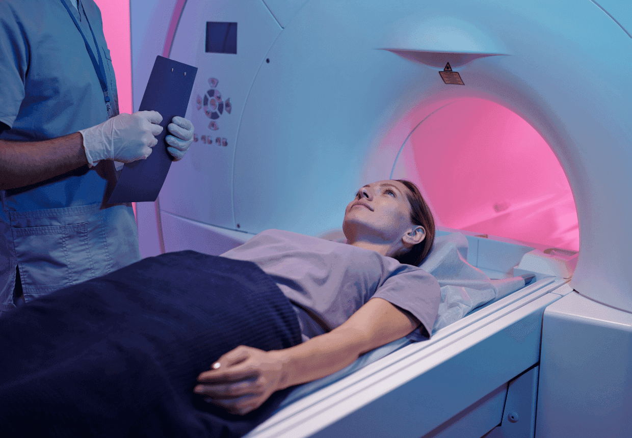
Generally speaking, people with modern dental implants can undergo MRI scans, but compatibility must be checked depending on the characteristics of the implant and the conditions of the examination. Dental implants are often made of titanium or ceramic materials, but the associated prosthetic components must also be taken into account. Before the scan, please inform the MRI team about any existing implants and dental work, as these may require additional checks or could affect the quality of the images.
In short:
- In most cases, modern dental implants do not prevent an MRI scan from being carried out, but safety must be confirmed for the specific device and the conditions of the scan.
- Titanium and zirconia implants are not strongly ferromagnetic.
- Dental work may cause artefacts, particularly during head and neck scans.
- Dentures with magnetic attachments require further checks.
- Do not remove a fixed dental restoration yourself before an MRI scan.
- Inform the imaging team of all existing implants and dental work.
Can you have an MRI scan if you have a dental implant?
Yes, in the vast majority of cases. The magnetic field of an MRI scanner strongly attracts only ferromagnetic materials, such as certain types of steel. Titanium and zirconia do not fall into this category. However, ‘non-magnetic’ does not mean ‘no interaction at all’: a metal may distort the image or heat up slightly. For this reason, medical devices are tested and labelled for specific examination conditions (so-called ‘MR Conditional’ devices).
To understand what components make up the implant and how it integrates into the bone, find out what a dental implant is and what the stages of treatment are.
Are titanium dental implants MRI-compatible?
Yes. Pure titanium and titanium alloys, including the titanium-zirconium alloy used in some implants, are not significantly attracted to a magnet. However, they can produce artefacts around the implant that are more extensive than those caused by ceramic implants.
Are zirconium dental implants compatible with MRI?
Ceramic implants made of zirconia (zirconium dioxide, a non-metallic material) interfere least with the MRI image of all the materials studied. However, some ceramic systems have metal screws or components, so the entire assembly is assessed.
Are there any risks to the dental implant during an MRI scan?
Patients are usually concerned about two things: that the implant might be ‘pulled’ by the magnet and that it might heat up. These are different phenomena. The attraction is due to the static magnetic field, whilst the heating is caused by the radio waves used to produce the image.
Is there a risk that the implant might move?
For a titanium or zirconium implant, the magnetic force is negligible, and the healed implant is integrated into the bone. Even with small ferromagnetic components fixed in the mouth, such as the plates of magnetic attachments, the forces measured remained well below the force with which they are bonded. The practical risk mainly concerns removable dentures with magnets.
Can the implant heat up during the scan?
The radio waves used during an MRI scan can cause implants and dental restorations containing metal to heat up. The extent of the heating depends on their shape and size, as well as on the MRI scanner and the settings used. In laboratory studies, the titanium mini-implants tested heated up by 0.35–0.47 °C, whilst bridges and a fixed full-arch restoration heated up by no more than 1.6–1.8 °C under the test conditions. These results relate to the specific items tested and cannot be generalised to all implants or dental restorations.
If you feel any heat or discomfort during the scan, please inform the MRI team immediately.
Can a dental implant affect MRI results?
It may affect image quality, though this does not mean that the scan is dangerous. Metal locally disrupts the magnetic field and produces an artefact: an area where the signal disappears or the image appears distorted. A systematic review published in 2022 identified artefacts of varying severity caused by metallic dental materials in MRI images of the head and neck; however, their diagnostic significance has not yet been established. The material, the amount of metal, the field strength and the sequences chosen all matter.
Brain or head and neck MRI with dental implants
This is where the impact is greatest. In the laboratory, the signal was affected up to approximately 1.4 cm around titanium implants, which may be significant when examining the oral cavity, the salivary glands or the temporomandibular joint. The radiologist can adjust the protocol if they know in advance what dental restorations you have.
MRI of the spine, abdomen or other body regions
When the area being examined is far from the mouth, the artefact caused by a dental implant does not usually appear in the image. It is still necessary to inform the team for safety reasons.
Can you have an MRI scan if you have crowns, bridges or other dental work?
Yes, but this is where the subtle distinction lies that many patients overlook. The implant is merely the artificial ‘root’ in the bone. On top of it sits the abutment and the prosthetic restoration (crown, bridge or denture), which may contain more metal than the implant itself. For an MRI scan, it is the whole assembly that matters.
‘I often see two opposing mistakes: the patient who doesn’t mention their dental work, because “they’re just teeth”, and the patient who is convinced they must remove their fixed restoration before an MRI scan. Neither is correct. Tell us about all the procedures and work you’ve had done, and the decision will be made by the imaging team and your dentist, based on the specific details.” – Dr. Rareș Nica, dentist, DENT ESTET
Metal-ceramic crowns, zirconia crowns and dental bridges
Metal-ceramic crowns have a metal framework, often made of chromium-cobalt or nickel-chromium alloys, which can produce greater artefacts than titanium. All-ceramic zirconia crowns generally cause minimal interference. Heat build-up in bridges is minimal. A fixed restoration should not be removed for an MRI scan without the dentist’s assessment.
Dental prostheses with magnetic retention systems
Specific measures are required here. The MRI field can demagnetise the magnets in the denture, causing it to lose its retention; therefore, the removable denture must be removed prior to the examination. The ferromagnetic plate remaining on the root or implant may produce a signal-free zone with a radius of up to 7–10 cm. For head scans, the dentist may temporarily remove the magnetic component screwed onto the implant, if requested by the imaging team. The retention is checked before and after the scan.
“Think of an implant restoration as a three-tiered structure. The first tier is the implant, the ‘root’ in the bone, usually made of titanium or, less commonly, zirconia. The second is the abutment, the part that connects to the visible tooth: it can be made of titanium or zirconia. The third is the restoration itself: the crown, bridge or denture, with a metal framework or made entirely of ceramic.
For an MRI scan, the right question is not just ‘what is the implant made of?’, but ‘how much metal do I have in my mouth, what type and where?’. A titanium implant matters little. A full-arch bridge with a cobalt-chromium framework or a magnetic prosthesis can count for much more, especially during a brain or head-and-neck MRI scan.
That is why I recommend that any patient with implants keep a simple record: the brand and serial number of the implants, the type of abutments and the material of the restoration. A few lines of information can save the imaging team from having to make assumptions.”
— Dr. Rareș Nica
| Type of restoration | Message for the patient |
|---|---|
| Dental implant made of titanium or a titanium alloy | Generally, it does not prevent an MRI scan from being carried out; it may cause artefacts around the implant, which are particularly relevant in head and neck examinations. |
| Zirconia ceramic implant | Generally, good compatibility and minimal interference with the image; any associated metal components should also be checked. |
| Metal-ceramic crown | May produce artefacts, particularly during head and neck examinations. |
| All-ceramic (zirconia) crown | Minimal interference with MRI imaging. |
| Fixed work on implants | The materials used in the prosthetic components are also important: the type of framework (titanium, cobalt-chromium, zirconia) and the amount of metal. |
| Denture with magnetic attachments | Requires specific assessment prior to an MRI scan; the removable denture is taken out, whilst the fixed components are assessed on a case-by-case basis. |
Find out more about zirconia dental crowns and the characteristics of this material used in dental restorations.
What should you do before an MRI scan if you have a dental implant?
Preparation is, first and foremost, about being well-informed. The steps below will help you arrive at your appointment with all the necessary information:
- Inform the imaging team about your implant and any dental work you have had, right from the time you book your appointment, not just in the questionnaire on the day of the examination.
- Provide the material, brand or model of the implant, if you know them. This information is usually recorded in your treatment record.
- Present the implant documentation, if available: the implant card or a medical letter.
- Mention any removable dentures, magnetic attachments and other implanted devices, including those outside the oral cavity.
- Follow the MRI team’s instructions regarding preparation for the scan.
The decision on whether to carry out the MRI and the conditions of the scan rests with the imaging team, who assess all devices present in the body. The dentist’s role is to provide accurate information about dental work and, if necessary, to remove any removable components. If you have any queries about your dental work, ask your dentist for a brief medical letter before your appointment.
MRI, CT or dental X-ray: what’s the difference?
These are different types of investigation, each with a different purpose. MRI provides excellent visualisation of soft tissues and does not use ionising radiation. However, for implant planning, the standard remains X-ray imaging: dental radiography and, when a three-dimensional assessment of the bone is required, computed tomography – abbreviated to CT or CBCT. Dental MRI is a promising area of research, currently used in selected situations rather than as a routine investigation. For a patient with implants, the choice of investigation depends on the clinical issue, not on the presence of the implant.
| Investigation | What needs to be explained |
|---|---|
| MRI | Uses a magnetic field and radio waves, without ionising radiation; particularly useful for soft tissues. |
| CT / CBCT | Uses X-rays; CBCT is frequently used in three-dimensional dental assessment and implant planning. |
| Dental X-ray | Uses X-rays for targeted assessments, such as checking an implant. |
Discover the differences between panoramic X-rays and dental CT scans, including their role in planning implant treatments.
Frequently asked questions about MRI and dental implants
Can I have an MRI scan straight after having a dental implant fitted?
Generally speaking, yes. The material of a titanium or zirconia implant is not attracted to a magnet, regardless of whether the bone has healed or not. Do, however, tell the team that you have recently had surgery: during a head scan, the post-operative swelling may be visible on the images.
Can I have a brain MRI if I have several dental implants?
Generally speaking, yes. More implants mean more metal and, potentially, more extensive artefacts in the lower part of the face. However, the brain is situated at a distance that usually limits the impact. The radiologist will choose the appropriate sequences if they are aware of this in advance.
Do I need to have my dental implant removed before an MRI scan?
No. The implant integrated into the bone does not need to be removed for an MRI scan. It may only be necessary for the dentist to remove the removable prosthesis or, in rare cases, to temporarily detach a screw-retained component, such as a magnetic attachment.
What should I do if I don’t know what material the dental implant is made of?
Contact the clinic where you had the treatment: the type of implant is usually recorded in your medical records. If this information cannot be obtained, tell the MRI team everything you know (the year of the procedure, the number of implants, the type of procedure), and they will decide how to proceed.
Can I have an MRI scan if I have a fixed bridge on several implants?
Generally speaking, yes. In full-arch restorations, the prosthetic framework may contain more metal than the implants, so it may produce more extensive artefacts. The heat build-up measured in the laboratory for such a restoration was low. Please inform the team of the material used for the prosthetic restoration.
Conclusion: a dental implant does not automatically constitute a contraindication for an MRI scan
Most modern dental implants do not prevent you from having an MRI scan. What you need to bear in mind is the effect that dental materials can have on the images, particularly during head and neck examinations, and in special cases, such as dentures with magnets. The most helpful thing you can do is to tell the imaging team about everything you have in your mouth.
Do you have any questions about the materials used in your implants or dental work? Talk to your dentist and ask for the necessary information before your MRI scan.